Vertebral pathological fractures and spinal instability due to metastases are a relevant clinical burden. Spinal Instability Neoplastic Score (SINS) proved useful to increase awareness of the risk among clinicians. To be actually predictive SINS would need to be improved through quantification of morphology, density, and biomechanics.
The Classify3D software, developed from the ALBA library, is a first step to the quantification of metastatic lesions through morphological analysis of CT images. The clinician is guided through simple steps to identify the affected vertebra, select two orthogonal sections, and trace the lesion borders. The software automatically estimates lesion volume, density and location within the vertebra. The software is being tested in an observational clinical study within the qSINS project.
Developed under the qSINS projectqSINS: quantitative revision of the Spinal Instability Neoplastic Score including CT-based assessment of spinal condition and finite element modeling of vertebral strength RF-2016-02364359
The following images describe a typical qSINS workflow to quantify the entity, density and intra-vertebral location of a metastatic lesion from CT or MRI data.
1) Importing CT/MRI patient images in DICOM format.

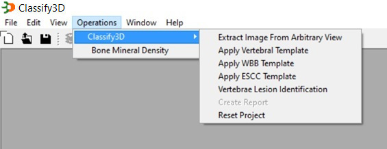
2) Upon selecting “Operation Menu”, two operations are available: a straightforward one to measure bone mineral density on a vertebral body, and the “Classify3D” to evaluate a lesion. These operations are available also through dedicated icons that, if navigated from left to right in the icons bar, define the workflow.
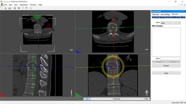
3) Defining the anatomical reference system: the operator defines the horizontal, sagittal and coronal axes for subsequent lesion tracing.
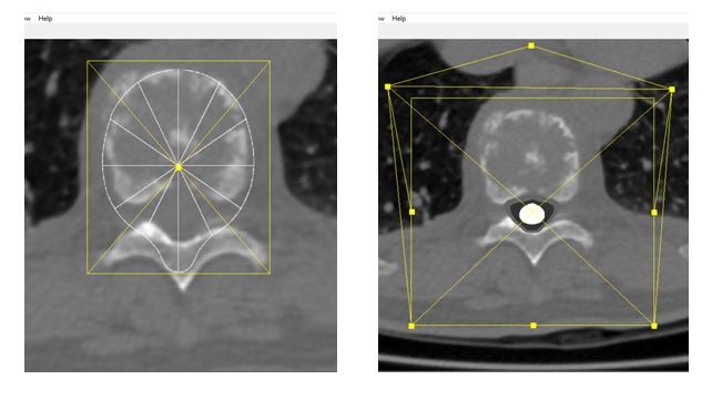
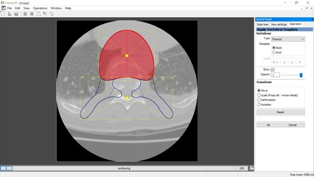
4) Selecting a vertebral template (thoracic, in this case) composed vertebral body and posterior vertebral arch parts.
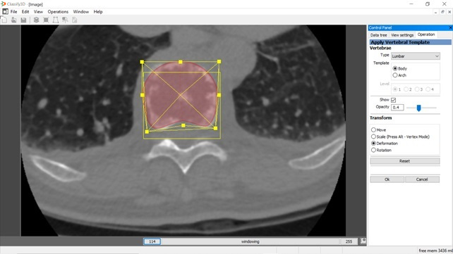
5) Morphing the template (vertebral body shown in this case), by moving, scaling, rotating and deforming the template.
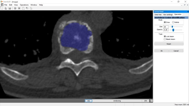
6) Segmenting the lesion on the horizontal section, with a customisable brush tool.
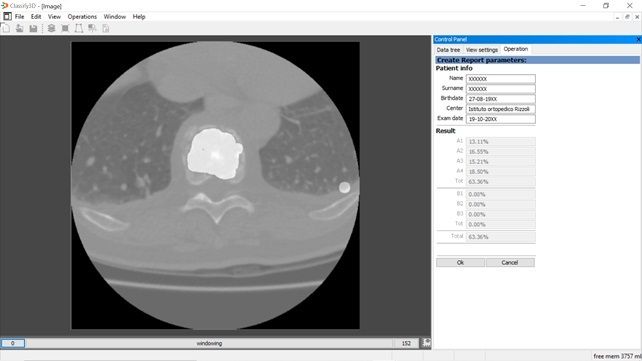
7) Computing the volumetric report: olume, relative volume, density, and location of the lesion within the vertebra are automatically computed and can be saved on a file.
8) (optional) Fitting additional templates for the WBB and ESCC classification oif the lesion.